The Complete Science of Visual Acuity: A Clinical Deep Dive
Visual acuity is the fundamental metric of optical clarity. From the historical foundations laid by Herman Snellen in 1862 to the modern pixel-perfect calibration algorithms used in this tool, we explore the physics, biology, and methodology behind how your eyes interpret the world.
The Fundamental Logic of the Snellen Line
A standard "20/20" letter is geometrically defined so that its total height subtends exactly 5 minutes of arc (5') at a distance of 20 feet. Furthermore, the thickness of the strokes and the gaps between them subtend exactly 1 minute of arc (1').
Chapter 1: The Evolution of the Optotype
Before the mid-19th century, vision testing was subjective. In 1862, Herman Snellen revolutionized the field by introducing the Optotype. Unlike standard typography, an optotype is designed on a strictly 5x5 grid.
The 5x5 Grid Architecture
Consider the Snellen 'E'. It consists of three horizontal arms and two vertical spaces. For an eye to resolve this as an 'E' rather than a black blur, the retina must be able to distinguish the white space from the black bar. Each bar and each space represents 1/5th of the total height. If the eye cannot resolve that 1 minute of arc gap, the letter merges into a block.
Chapter 2: Deciphering the Snellen Fraction
The vision score (e.g., 20/40 or 6/12 in metric) is a comparison ratio, not a percentage.
- 20/20 (6/6): You can see at 20 feet what a "standard" observer sees at 20 feet. This is normal, but not "perfect."
- 20/40 (6/12): You must be at 20 feet to see what a normal person sees at 40 feet.
- 20/200 (6/60): Legal blindness threshold in the US (best corrected).
Chapter 3: Anatomical Factors Limiting Acuity
Why can't we see infinitely small details? The limit is physical, biological, and neurological.
1. Cone Density in the Fovea

The fovea centralis is a tiny pit in the center of the macula responsible for sharp central vision. It contains the highest density of cone photoreceptors. The average distance between cone centers is about 2.5 micrometers (approx 0.5 minutes of arc). This creates a "sampling limit"—if two points of light fall on a single cone, they cannot be distinguished as two separate points.
2. Diffraction and the Airy Disk
Even with perfect optics, light passing through the pupil diffracts. A point source of light doesn't focus to a perfect point on the retina but forms a bullseye pattern called an Airy Disk. If the pupil is too small, diffraction increases, blurring the image. If the pupil is too large, spherical aberrations increase. The "sweet spot" for maximum acuity is generally a pupil diameter of 3mm to 4mm.
Chapter 4: Refractive Errors Explained
When you cannot read the bottom line of this chart, it is usually due to a Refractive Error—a mismatch between the eye's length and its optical power.
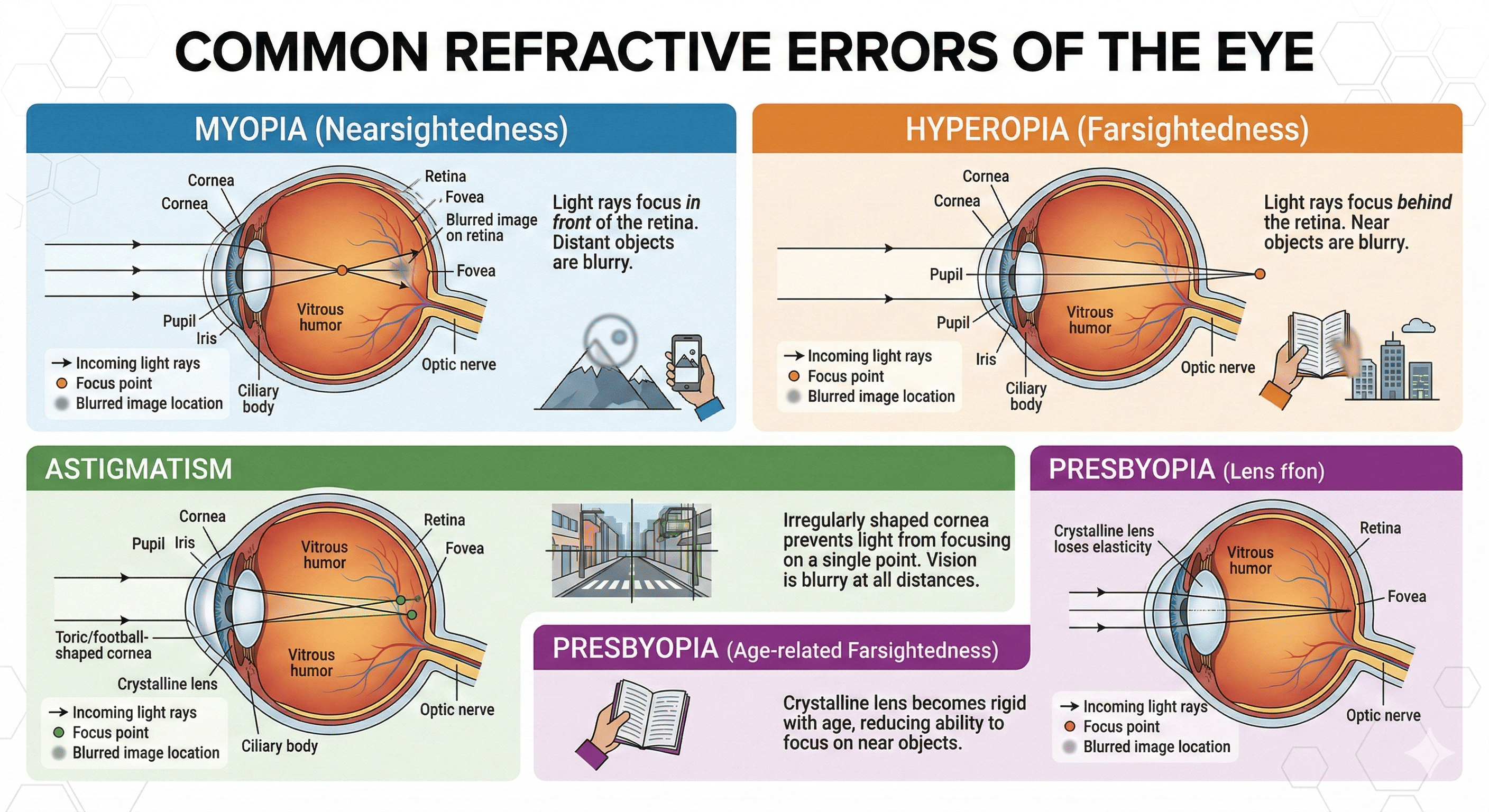
Myopia (Nearsightedness)
The Mechanics: The eyeball is too long (axial myopia) or the cornea is too
steep. Light rays focus at a point in front of the retina. By the time they hit the
retina, they have diverged, creating a blur circle.
The Experience: Near objects are clear; distant objects are blurry. This is the
most common refractive error globally, often exacerbated by lack of sunlight exposure during
childhood.
Hyperopia (Farsightedness)
The Mechanics: The eyeball is too short, or the cornea is too flat. Light
attempts to focus at a theoretical point behind the retina.
The Experience: Young people can often "accommodate" (flex the ciliary muscle
to thicken the lens) to bring the focal point forward. However, this causes significant eye
strain, headaches, and difficulty sustaining near focus.
Astigmatism
The Mechanics: The cornea or lens is not perfectly spherical (like a basketball)
but toric (like an American football). This creates two different focal points perpendicular to
each other.
The Experience: Vision is distorted or shadowed at all distances. An 'E' might
look like it has a ghost image hovering slightly above it.
Chapter 5: Pediatric Vision Screening
Vision screening in children is critical because the visual pathway between the eye and the brain is still developing until around age 7-9. If a child has a strong refractive error in one eye, or a strabismus (eye turn), the brain may ignore input from that eye to prevent double vision.
This leads to Amblyopia (Lazy Eye). If not caught early, the vision loss becomes permanent, as the neurological connections atrophy. This is why the "Tumbling E" chart (available in this tool) is vital—it allows testing of children who do not yet know the alphabet.
Chapter 6: Nutrition and Ocular Health
While carrots (Vitamin A) are famous for eye health, modern research highlights other critical nutrients for maintaining acuity and preventing macular degeneration:
- Lutein and Zeaxanthin: Found in dark leafy greens (spinach, kale), these carotenoids act as "internal sunglasses," filtering out harmful high-energy blue light and protecting the macula.
- Omega-3 Fatty Acids: Found in fatty fish, these support the structural integrity of the retina and the meibomian glands that produce the oily layer of tears, preventing dry eye.
- Zinc: High concentrations of zinc are found in the eye; it helps transport Vitamin A from the liver to the retina to produce melanin, a protective pigment.
Chapter 7: The 20-20-20 Rule & Digital Strain
In our digital era, Computer Vision Syndrome is prevalent. The ciliary muscle must remain contracted to focus on near screens. To prevent spasm and strain, the American Optometric Association recommends the 20-20-20 rule:
- Every 20 minutes...
- Look at something 20 feet away...
- For at least 20 seconds.
This tool helps facilitate that. By setting the chart to 3m or 6m, you are forcing your eyes to relax their accommodation and focus at a distance.
Chapter 8: The Physics of the Minimal Angle of Resolution (MAR)
While the Snellen fraction is the household name for vision, clinicians often use the LogMAR scale for scientific accuracy. The Minimal Angle of Resolution (MAR) is the smallest spatial gap a person can resolve, measured in minutes of arc.
The relationship between a Snellen fraction and MAR is inverse:
Example: For 20/40 vision, the MAR is $40/20 = 2$ minutes of arc. This means the viewer can only resolve details twice as large as a standard 20/20 observer.
Chapter 9: Contrast Sensitivity — Beyond the Black & White
Visual acuity is often tested in high-contrast environments (black letters on a white background). However, real-world "functional vision" relies on Contrast Sensitivity. This determines your ability to distinguish an object from its background, such as seeing a gray car on a foggy highway or steps in a dimly lit hallway.
- Glare Disability: Light scattering within the eye (often from cataracts) can "wash out" contrast even if your Snellen acuity remains 20/20.
- Spatial Frequency: This measures how well you see alternating light and dark bands. Low spatial frequency helps you see large objects (like a face or a doorway), while high spatial frequency is needed for resolving fine details (like reading small print).
- Testing Tools: While the Snellen chart tests high-contrast acuity, specialized charts test contrast sensitivity using letters of the same size but decreasing contrast.
Chapter 10: The Inevitability of Presbyopia
Often confused with hyperopia (farsightedness), Presbyopia is an age-related condition that eventually affects everyone, usually beginning in the early to mid-40s. It is a highly searched topic as individuals notice a sudden change in their near-vision capabilities.
The Loss of Accommodation
Inside the eye, the crystalline lens changes shape to focus on near objects—a process called accommodation. In youth, this lens is highly elastic. As we age, the lens proteins change, making it thicker and more rigid. Simultaneously, the ciliary muscles may lose some of their functional elasticity.
When the lens can no longer flex sufficiently to increase its optical power, near objects become blurry. This is the physiological reason why people begin holding reading material at arm's length. It is not a disease, but a natural evolution of the ocular anatomy, typically corrected with reading glasses, bifocals, or progressive lenses.
Frequently Asked Questions (Clinical & Functional)
Optimized for quick answers to common clinical queries regarding visual acuity and eye health.
Is 20/15 vision better than 20/20?
Yes. If you have 20/15 vision, it means you can see at 20 feet what a person with normal (20/20) vision can only see if they step closer to 15 feet. Human maximum visual acuity is biologically limited to roughly 20/10 or 20/8 due to the density of photoreceptors in the fovea.
Can you naturally improve your visual acuity?
Structural refractive errors (myopia, hyperopia, astigmatism) cannot be cured with "eye exercises" or diet. However, maintaining a diet rich in Lutein, Zeaxanthin, and Omega-3 fatty acids can prevent macular degeneration and dry eye, preserving your baseline acuity as you age.
What does "Legally Blind" mean in terms of visual acuity?
In the United States, legal blindness is strictly defined as a visual acuity of 20/200 or worse in the better-seeing eye with the best possible optical correction (glasses or contacts), or a visual field restricted to 20 degrees or less.
How often should adults get a comprehensive eye exam?
The American Optometric Association recommends comprehensive eye exams every two years for asymptomatic adults aged 18 to 64. Annual exams are recommended for adults 65 and older, or anyone with diabetes, hypertension, or a family history of ocular disease.
What is contrast sensitivity and why is it important in vision tests?
Contrast sensitivity measures your ability to distinguish an object from its background. Testing with lower contrast levels helps identify early signs of conditions like cataracts or glaucoma, even if your standard Snellen visual acuity is perfectly 20/20.
Protect Your Sight
High-precision screening is the first step toward long-term ocular health.
Restart Calibration